Know the Different Types of Echocardiogram Tests
Introduction
Discovering the inner workings of our hearts has always been a marvel, and Echocardiogram serves as a window into this intricate world. Imagine peering into the chambers of your heart with the clarity of a crystal-clear mirror. Echocardiogram, often simply called an “echo,” encompasses various types, each offering a unique perspective on cardiac health. From the gentle touch of a traditional echocardiogram to the advanced insights of transesophageal Echocardiogram (TEE), these techniques reveal the heartbeat’s secrets with breathtaking clarity. Join us as we delve into the fascinating world of cardiac imaging and learn how different types of Echocardiogram contribute to a better understanding of heart conditions.
What is Echocardiogram?
Echocardiogram, often referred to simply as an “echo,” is a non-invasive imaging procedure that utilizes sound waves to create real-time images of the heart. Similar to ultrasound imaging used in prenatal care, Echocardiogram focuses specifically on the heart’s chambers, valves, and blood flow patterns. By providing detailed images of the heart’s anatomy and function, Echocardiogram enables healthcare professionals to diagnose a wide range of cardiac conditions.
The images can also help them get information
Echocardiographic images offer clinicians a wealth of information, allowing them to assess the size and thickness of the heart chambers, evaluate the integrity and function of heart valves, detect abnormalities such as blood clots or tumors, and assess blood flow dynamics within the heart and major blood vessels.
Why is an Echocardiogram Performed?
Echocardiogram serves multiple purposes in clinical practice:
- Diagnosis: It helps diagnose various heart conditions such as heart valve diseases, congenital heart defects, cardiomyopathies, and pericardial diseases.
- Assessment of Function: Echocardiogram assesses the heart’s pumping function, detecting abnormalities in its contractility and overall performance.
- Monitoring: It aids in monitoring the progression of heart diseases and the effectiveness of treatments over time.
- Guidance: Echocardiogram guides procedures such as cardiac catheterization and valve replacement surgeries by providing real-time imaging.
Explore Different Echocardiogram Types
Transthoracic Echocardiogram
Transthoracic Echocardiogram (TTE), also known as two-dimensional Echocardiogram (2D echo), is the most common and widely used type of Echocardiogram. It provides valuable information about the overall structure and function of the heart.
During a TTE procedure, a transducer is placed on the chest wall to capture high-resolution images of the heart. This non-invasive technique allows healthcare professionals to assess the size, shape, and movement of the heart chambers, as well as the thickness and function of the heart muscle.
TTE is often used to diagnose and monitor a wide range of heart conditions, including heart valve abnormalities, heart failure, congenital heart defects, and diseases of the heart muscle. It provides real-time visualization, allowing healthcare providers to evaluate the blood flow through the heart and detect any abnormalities or obstructions.
In addition to its diagnostic capabilities, TTE is also a valuable tool for guiding various cardiac procedures, such as the placement of cardiac devices or the monitoring of interventions. Its wide availability, safety, and non-invasive nature make TTE an essential component of comprehensive cardiac imaging.
Overall, transthoracic Echocardiogram (TTE) plays a critical role in evaluating heart health by providing detailed and accurate imaging of the heart’s structure and function. Its widespread use and effectiveness make it an indispensable tool in the field of cardiology.
Transesophageal Echocardiogram

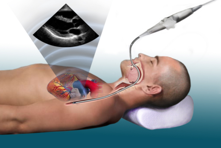
Transesophageal Echocardiogram (TEE) is a more invasive type of Echocardiogram that involves inserting a probe through the mouth and down into the esophagus. This allows for close-up and detailed imaging of the heart structures that may not be visible with transthoracic Echocardiogram (TTE).
TEE is commonly used during surgeries or when a more detailed evaluation is required. It provides healthcare professionals with valuable insights into heart conditions by capturing high-resolution images of the heart’s internal structures.
By inserting the probe into the esophagus, TEE eliminates the barrier created by the ribcage and lungs, allowing for clearer visualization of the heart. This technique can detect various cardiac abnormalities, such as blood clots, valve malfunctions, and structural defects.
During the procedure, the patient is usually given a local anaesthetic to numb the throat and may receive sedation to ensure comfort. Once the probe is in position, the sonographer or cardiologist will manoeuvre it to obtain images from different angles.
TEE can also be used to guide other procedures, such as transcatheter interventions or surgeries, providing real-time imaging guidance for the accurate placement of devices or instruments. In summary, transesophageal Echocardiogram plays a crucial role in diagnosing and monitoring heart conditions by offering detailed and up-close images of the heart structures. As a more invasive technique, TEE allows healthcare professionals to obtain valuable insights for effective treatment planning and improved patient outcomes.
Stress Echocardiogram
Stress Echocardiogram, also known as exercise Echocardiogram, is a powerful technique that combines Echocardiogram with exercises or medications that increase the heart rate. By subjecting the heart to stress, this procedure helps evaluate its response and identifies any abnormalities in blood flow or heart function that may not be visible at rest. Stress Echocardiogram offers valuable insights into the heart’s performance during physical exertion, allowing healthcare professionals to diagnose and monitor cardiac conditions more effectively.
Doppler Echocardiogram
Doppler Echocardiogram is a valuable tool in assessing blood flow and velocity within the heart. By utilizing the Doppler effect, this technique provides crucial information about the direction, speed, and volume of blood flow. Doppler Echocardiogram can be used in conjunction with other types of Echocardiogram, such as transthoracic Echocardiogram (TTE) or transesophageal Echocardiogram (TEE), to enhance the evaluation of cardiac function and diagnose various heart conditions.
Three-dimensional Echocardiogram
Three-dimensional Echocardiogram is an advanced imaging technique that provides a detailed, real-time visualization of the heart in three dimensions. Unlike traditional two-dimensional Echocardiogram, which offers a flat image of the heart, 3D Echocardiogram captures a more realistic representation of cardiac structures, allowing for a comprehensive assessment of the heart’s anatomy and function.
This technology utilizes specialized ultrasound probes and advanced software algorithms to construct detailed 3D images, offering healthcare professionals a clearer understanding of complex cardiac structures such as heart valves, chambers, and vessels. A three-dimensional Echocardiogram is particularly useful in evaluating congenital heart defects, assessing valve function, and planning complex cardiac procedures, making it a valuable tool in modern cardiology practice.
Echocardiogram Procedure
During an echocardiogram, the patient lies on a table while a trained technologist places a transducer on various areas of the chest wall. A gel is applied to improve sound wave transmission. The transducer emits high-frequency sound waves, which bounce off cardiac structures and are captured by the transducer, creating real-time images on a monitor. The procedure is painless and typically takes 30 to 60 minutes to complete.
What Results Will I Receive with Echocardiogram Reports?
After the echocardiogram, a cardiologist interprets the images and generates a report detailing the heart’s structure, function, and any abnormalities detected. Depending on the findings, further diagnostic tests or treatments may be recommended. Patients are often briefed on their results during a follow-up appointment with their healthcare provider.
Conclusion
An echocardiogram represents a cornerstone in the diagnosis and management of cardiovascular diseases, offering a safe, non-invasive, and highly informative imaging modality. With its various types tailored to specific clinical scenarios, Echocardiogram continues to play a pivotal role in improving patient outcomes and enhancing our understanding of cardiac pathophysiology. As technology advances further, the future of Echocardiograms holds promise for even greater precision and diagnostic capabilities, ushering in a new era of cardiac care.
Get Your Echocardiogram Test Done at Precision Pathlab
Get your Echocardiogram done at Precision Pathlab, the best diagnostic center in Jaipur. Our state-of-the-art facility offers top-notch Echocardiogram services conducted by experienced professionals. Whether you need a routine heart assessment or a comprehensive evaluation of cardiac function, we ensure accurate and reliable results to guide your healthcare journey. Moreover, Precision Pathlab also provides full body checkup in Jaipur, catering to your overall health needs. With our commitment to excellence and patient-centric approach, you can trust us to deliver exceptional diagnostic services for your peace of mind and well-being.